Jump to content

File:Stomach mucosal layer labeled.svg

Page contents not supported in other languages.
This is a file from the Wikimedia Commons
From Wikipedia, the free encyclopedia

Original file (SVG file, nominally 637 × 634 pixels, file size: 284 KB)

Summary

Description
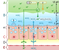
English: Diagram of alkaline Mucous layer in stomach with mucosal defense mechanisms
Polski: Schemat budowy błony śluzowej żołądka z uwzględnieniem mechanizmów bariery śluzowo-alkalicznej
日本語: 粘膜防御機構を備えた胃の塩基性粘液層の図
 
This W3C-unspecified vector image was created with Inkscape .
Date
Source

Own work using:

  1. Fr. Boumphrey, File:Stomach mucous.png
  2. S.J. Konturek, P.C. Konturek, T. Pawlik, Z. Sliwowski, W. Ochmanski, E.G. Hahn, Duodenal mucosal protection by bicarbonate secretion and its mechanisms [1]
  3. John T. Hansen, PhD and Bruce M. Koeppen, MD, PhD, Netter's Atlas of Human Physiology Mucosal Defense Mechanisms
Author

MKomorniczak -talk- 11:43, 23 April 2011 (UTC)

Illustration by : Michał Komorniczak
This file has been released into the Creative Commons 3.0. Attribution-ShareAlike (CC BY-SA 3.0)

If you use on your website or in your publication my images (either original or modified), you are requested to give me details: Michał Komorniczak (Poland) or Michal Komorniczak (Poland).

For more information, write to my e-mail address: m.komorniczak.pl@gmail.com

Permission
(Reusing this file)
I, the copyright holder of this work, hereby publish it under the following license:
w:en:Creative Commons
attribution
This file is licensed under the Creative Commons Attribution 3.0 Unported license.
You are free:
  • to share – to copy, distribute and transmit the work
  • to remix – to adapt the work
Under the following conditions:
  • attribution – You must give appropriate credit, provide a link to the license, and indicate if changes were made. You may do so in any reasonable manner, but not in any way that suggests the licensor endorses you or your use.
Other versions

[edit]

Label

Label PL - polski EN - english
A Światło żołądka Gastric lumen
1 Pepsynogen Pepsinogen
2 Pepsyna Pepsin
B Warstwa śluzowa Mucus-bicarbonate Barrier
3 śluz Mucus
C Komórki nabłonkowe powierzchowne Epithelial Tight Junctions
D Ścisłe połączenie międzykomórkowe Surface Mucous Cells
E Substancja międzykomórkowa Interstitial Fluid
F Naczynie włosowate Capillary

This image has been assessed using the Quality image guidelines and is considered a Quality image.

العربية  جازايرية  беларуская  беларуская (тарашкевіца)  български  বাংলা  català  čeština  Cymraeg  Deutsch  Schweizer Hochdeutsch  Zazaki  Ελληνικά  English  Esperanto  español  eesti  euskara  فارسی  suomi  français  galego  עברית  हिन्दी  hrvatski  magyar  հայերեն  Bahasa Indonesia  italiano  日本語  Jawa  ქართული  кыргызча  한국어  kurdî  Lëtzebuergesch  lietuvių  македонски  മലയാളം  मराठी  Bahasa Melayu  Nederlands  Norfuk / Pitkern  polski  português  português do Brasil  qaraqalpaqsha  rumantsch  română  русский  sicilianu  slovenčina  slovenščina  shqip  српски / srpski  svenska  தமிழ்  తెలుగు  ไทย  Tagalog  toki pona  Türkçe  українська  Oʻzbekcha  vèneto  Tiếng Việt  中文  中文(简体)  中文(繁體)  +/−

Captions

Diagram of alkaline Mucous layer in stomach with mucosal defense mechanisms

Items portrayed in this file

depicts

23 April 2011

File history

Click on a date/time to view the file as it appeared at that time.

Date/TimeThumbnailDimensionsUserComment
current11:51, 26 August 2011Thumbnail for version as of 11:51, 26 August 2011637 × 634 (284 KB)M.Komorniczakph - shadow
11:50, 26 August 2011Thumbnail for version as of 11:50, 26 August 2011637 × 634 (284 KB)M.Komorniczaksmall fix
11:46, 26 August 2011Thumbnail for version as of 11:46, 26 August 2011637 × 634 (284 KB)M.Komorniczaksame fix
11:36, 26 August 2011Thumbnail for version as of 11:36, 26 August 2011637 × 634 (281 KB)M.Komorniczakbiger size
13:39, 23 August 2011Thumbnail for version as of 13:39, 23 August 2011396 × 387 (279 KB)M.Komorniczakzmiana wyglądu
11:56, 23 April 2011Thumbnail for version as of 11:56, 23 April 2011409 × 351 (155 KB)M.Komorniczakvector.
11:43, 23 April 2011Thumbnail for version as of 11:43, 23 April 2011409 × 351 (155 KB)M.Komorniczak{{Information |Description= |Source={{own}}, based in the information and diagrams found in: # Fr. Boumphrey, File:Stomach mucous.png # S.J. Konturek, P.C. Konturek, T. Pawlik, Z. Sliwowski, W. Ochmanski, E.G. Hahn, Duodenal mucosal protection by bic

The following 3 pages use this file:

Global file usage

The following other wikis use this file: